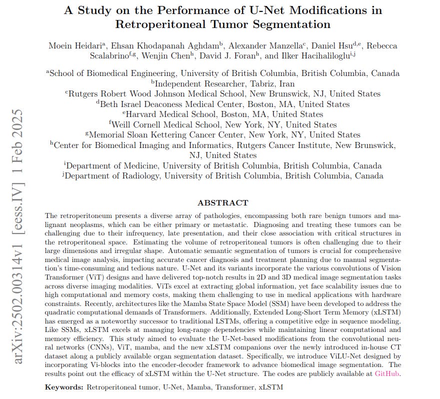
"A Study on the Performance of U-Net Modifications in Retroperitoneal Tumor Segmentation"
Below podcast on this paper is generated with Google's Illuminate.
https://arxiv.org/abs/2502.00314
Retroperitoneal tumors pose diagnostic and treatment challenges due to their complex location and irregular shapes, making manual volume estimation and segmentation difficult and time-consuming. This paper introduces ViLU-Net, a U-Net-based architecture incorporating Vision xLSTM (ViL) blocks, to improve automatic retroperitoneal tumor segmentation.
ViLU-Net leverages xLSTM's efficiency in handling long-range dependencies within medical images, offering a computationally effective alternative to Transformer-based methods.
-----
📌 ViLU-Net effectively replaces computationally heavy Transformers with efficient xLSTM blocks within the U-Net. This maintains segmentation accuracy while reducing computational cost. It makes high-resolution medical image analysis more practical.
📌 The ViLU-Net architecture leverages the strengths of both CNNs and xLSTMs. CNNs capture local details, while xLSTMs model long-range dependencies. This hybrid approach enhances segmentation performance in complex medical images.
📌 By achieving superior Dice Similarity Coefficient scores (0.9309 on tumors), ViLU-Net directly improves diagnostic precision. Accurate tumor segmentation leads to better treatment planning and monitoring for retroperitoneal cancers.
----------
Methods Explored in this Paper 🔧:
→ The paper introduces ViLU-Net, a modified U-Net architecture for medical image segmentation.
→ ViLU-Net incorporates Vision xLSTM (ViL) blocks within its encoder and decoder.
→ ViL blocks use modified Long Short-Term Memory (mLSTM) layers to capture spatial and temporal dependencies efficiently.
→ The architecture starts with a convolutional stem for initial feature extraction.
→ It uses an encoder-decoder structure with skip connections, similar to U-Net, but replaces standard convolutional layers with ViL blocks.
→ mLSTM blocks in ViL are designed for parallel processing and feature a matrix memory with covariance updates.
→ Odd and even numbered mLSTM blocks process patch tokens in opposite directions to enhance feature representation.
-----
Key Insights 💡:
→ xLSTM based architectures can offer improved accuracy and efficiency for medical image segmentation.
→ ViLU-Net effectively combines Convolutional Neural Networks' (CNNs) local feature extraction with xLSTM's long-range dependency modeling.
→ The proposed ViLU-Net architecture demonstrates superior or comparable performance to state-of-the-art methods like Transformer-based and Mamba-based U-Nets.
→ xLSTM presents a computationally efficient alternative to Transformers for medical imaging tasks, especially in resource-constrained environments.
-----
Results 📊:
→ On the abdomen CT dataset, ViLU-Net achieved a Dice Similarity Coefficient (DSC) of 0.8594, outperforming nnU-Net (0.8469), SwinUNETR (0.8259), and U-Mamba (0.8480).
→ On the retroperitoneal tumor dataset, ViLU-Net achieved a DSC of 0.9309, Normalized Surface Distance (NSD) of 0.9292, Hausdorff Distance (HD) of 11.19, and Intersection over Union (IoU) of 0.8720.
→ ViLU-Net showed qualitative improvements in tumor boundary delineation and reduced false positives compared to other models on the retroperitoneal tumor dataset.


